تخطي للذهاب إلى المحتوى

Empower Your Practice

Upskill with NTVet Online Courses

Advance Your Veterinary Knowledge & Skills with NTVet Online Courses

لوحة الصدارة

لا توجد لوحة صدارة في الوقت الحالي :(

عرض الكل
الدورات الأكثر شعبية

أكلاريكس LX9 الموجات فوق الصوتية

1. The Diagnostic Ultrasound System consists of a main system and associated ultrasound transducers.

2. The system circuitry generates an electronic voltage pulse, which is transmitted to the transducer. In the transducer, a piezoelectric array converts the electronic pulse into an ultrasonic pressure wave. When coupled to the body, the pressure wave transmits through body tissues. The waves are then reflected within the body and detected by the transducer, which then converts the waves back to an electrical signal. The system then analyzes the returned signals and generates an ultrasound image or spectral Doppler display.

3. The Diagnostic Ultrasound System provides the operator the ability to measure anatomical structures, and offers analysis packages that provide information used by competent health care professionals to make a diagnosis.

4. The system‟s user interface provides both hard keys for functions frequently used throughout an exam and touch screen controls for mode-specific functions.

EDAN اجهزة
أكلاريكس LX3 الموجات فوق الصوتية

1. The Acclarix LX3 series Diagnostic Ultrasound System consists of a main system and associated

ultrasound transducers.

2. The system circuitry generates an electronic voltage pulse, which is transmitted to the transducer. In

the transducer, a piezoelectric array converts the electronic pulse into an ultrasonic pressure wave.

When coupled to the body, the pressure wave transmits through body tissues. The waves are then

reflected within the body and detected by the transducer, which then converts the waves back to an

electrical signal. The system then analyzes the returned signals and generates an ultrasound image

or spectral Doppler display.

3. The Diagnostic Ultrasound System provides the operator the ability to measure anatomical structures,

and offers analysis packages that provide information used by competent health care professionals to

make a diagnosis.

4. The system’s user interface provides both hard keys for functions frequently used throughout an exam

and touch screen controls for mode-specific functions.

EDAN اجهزة
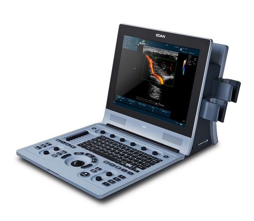
U60 VET الموجات فوق الصوتية البيطرية

The U60 VET is a portable Veterinary Diagnostic Ultrasound System, which applies advanced

technologies such as Phased Inversion Harmonic Compound Imaging (eHCI),

Multi-Beam-Forming (mBeam), Speckle Resistance Imaging (eSRI), and Spatial Compounding

Imaging, etc. Various image parameter adjustments, 15 inch LCD and diverse probes are

configured to provide clear and stable images.

Essential Performance:

Satisfying 2D imaging and basic functions of color ultrasonic system (such as color flow imaging

and spectral Doppler imaging).

1. 2D Imaging: It provides B mode and M mode imaging.

2. Color flow imaging.

3. Spectral Doppler imaging.

EDAN اجهزة
عرض الكل
أحدث الدورات

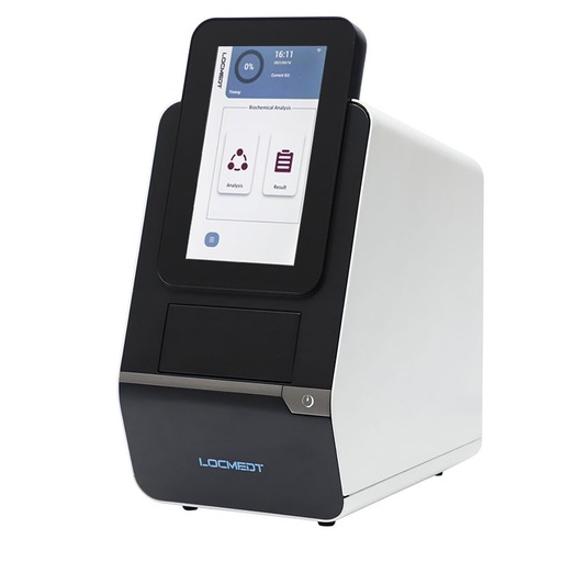
محلل الكيمياء الحيوية الأوتوماتيكي LOC-200

LOC-200 Automatic Biochemistry Analyzer is designed to perform the blood biochemistry tests and electrolyte analysis, delivering reliable and accurate results around 8 minutes by 3 drops of blood sample (90-120μL).

Orthopedic Plates
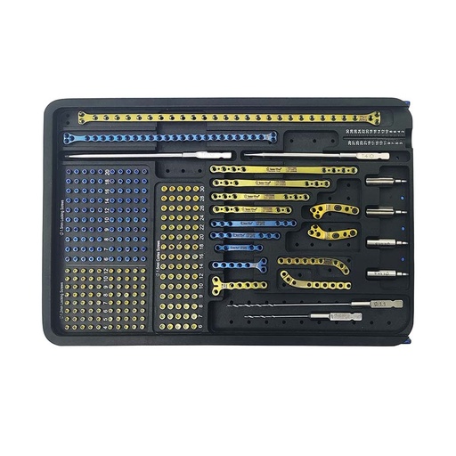
Orthopedic Plates

An orthopedic plate is a form of internal fixation used in orthopaedic surgery to hold fractures in place to allow bone healing and to reduce the possibility of nonunion. Most modern plates include bone screws to help the orthopedic plate stay in place.
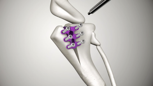
Titanium TTA Cages (تقدم الحدبة الظنبوبية) - عده

The TTA Cage serves the role of bone spacer that is placed between the two dissected section of the tibia. It has two slots for two bone screws which are tightly inserted into bone going through the slots. This holds the dissected bone together increasing the angle on the relationship between the femur and the tibia.